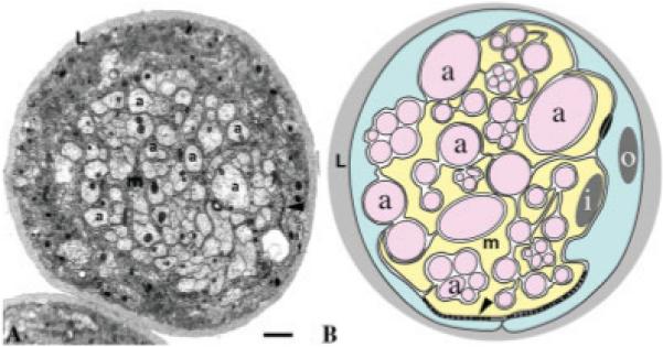
Fig. 2.

Peripheral nerve insulation in Drosophila. A: Transverse section through a Drosophila larval peripheral nerve showing individual axons (a) being wrapped by the membrane (m) of the inner glial cell. This in turn is wrapped by another glial cell, the perineurial glial cell. Extensive SJs were established between perineurial and inner glial membranes (arrowhead; Banerjee et al., 2006a). There is an outer-most layer of neural lamella (L) surrounding the nerve fibers. Scale bar = 1 μm. B: Schematic of a Drosophila peripheral nerve showing the axons and fascicles (a) surrounded by a sheath formed by the inner ensheathing glial cell (i), which in turn is surrounded by the outer perineurial glial cells (o). Arrowhead points to the areas where septate junctions formed between glial membranes.
