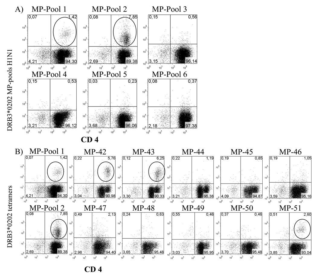
Figure 3. Identification of DRB3*0202-restricted MP specific epitopes.
A) Representative staining profiles with DRB3*0202-MP tetramer pools. CD4+ T cells from a DRB3*0202 subject were stimulated with six pools of MP peptides, and stained with DRB3*0202 pooled peptide tetramers 14 days after stimulation. Circled events indicate populations that were tetramer positive. B) Fine mapping was performed for the tetramer positive pools (MP pool 1 and MP pool 2) using individual peptide tetramers. MP1–20 (MP42), MP9–28 (MP43), and MP73–92 (MP51) were identified as peptides that contain DRB3*0202 restricted epitopes, negative individual peptide tetramers from MP pools 1 and 2 are also represented. TGEM with MP pools was performed in two individuals.

