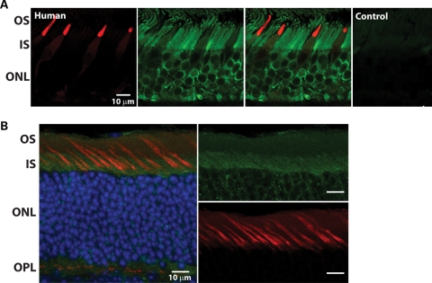
Figure 1.
AIPL1 is expressed in adult cone photoreceptors. (A) Immunolocalization of human-specific AIPL1 antibody (green) to the outer nuclear layer (ONL) of human retina. Anti-AIPL1 labeled rods intensely from the synaptic terminals to the proximal outer segment, extending along the axoneme. Cones, identified by mAb 7G6 labeling (red), appeared weakly labeled in the myoid and ellipsoid. Sections with omission of both primary antibodies served as negative controls. OS, outer segment; IS, inner segment. Scale bars, 10 µm. (B) AIPL1 (green) is expressed in both rod and cone photoreceptors of C57/BL6 mouse retina (post-natal day 135). Colocalization (yellow) of mAIPL1 immunoreactivity and peanut agglutinin (red), a cone marker, suggests that AIPL1 is expressed in mouse cones. ONL nuclei are visualized by DAPI staining (blue). OPL, outer plexiform layer. Scale bars, 10 µm.

