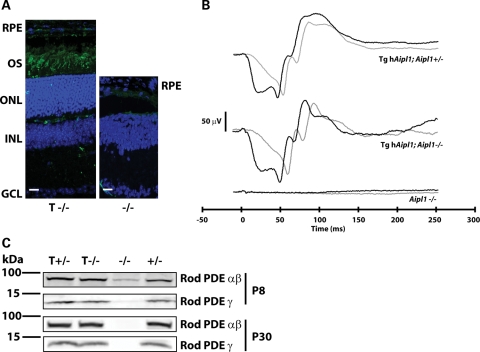
Figure 3.
Transgene expression of human AIPL1 restores rod morphology and function. (A) Expression of hAIPL1 in rod photoreceptors abrogates the rod cell death observed in Aipl1−/− mice. Rod outer segments (OS) were labeled with anti-rod opsin antibody (4D2, green) and the outer nuclear layer (ONL) was visualized with DAPI (blue). In tg hAipl1; Aipl1−/−, the ONL was recovered to near normal thickness (8–10 nuclear layers) and rod opsin is clearly visible in the ROS (left panel). However, at the same age, degeneration of the ONL is complete and no rod opsin can be detected in the retina of Aipl1−/− mice (right panel). Sections are taken from P100 mice. RPE, retinal pigment epithelium; OS, outer segments; ONL, outer nuclear layer; INL, inner nuclear layer; GCL, ganglion cell layer. Scale bars, 20 µm. (B) Full-field dark-adapted ERG recording from P15 mice show complete recovery of rod light response. Tg hAipl1; Aipl1+/− mice exhibit robust rod-derived electrical responses that are reciprocated in tg hAipl1; Aipl1−/− mice. This is in contrast to Aipl1−/− mice which exhibit no light-driven rod photoreceptor response. Black lines represent a light intensity of 2.5 cd s m−2 and gray lines represent a lower light intensity of 0.16 cd s m−2. These recordings are an average of at least three different mice. Littermates were used to compare responses. (C) Recovery of rod PDE6 expression. Using MOE antibody (1:2000), rod PDE6 can be detected in Aipl1+/− (+/−), tg hAipl1; Aipl1+/− (T +/−) and tg hAipl1; Aipl1−/− (T −/−) mice as early as P8. However, rod PDE6 levels are reduced in Aipl1−/− (−/−) mice at P8 and undetectable at P30.

