Figure 5.
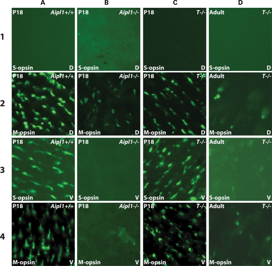
Cone degeneration is slow in the presence of rescued rod photoreceptors. Whole-mounted retinas from P18 Aipl1+/+ (A), Aipl1−/− (B), tg hAipl1; Aipl1−/− (T−/−) (C) and adult (P40) tg hAipl1; Aipl1−/− (D) were labeled with anti-S- and M-opsin antibodies. Images were taken from the dorsal (D) and ventral (V) regions of the retina. Aipl1+/+ retina (A) shows typical distribution of S-opsin containing cones in the ventral retina (A3) and M-opsin containing cones throughout the dorsal and ventral retina (A2 and A4). The majority of cone photoreceptors degenerate by P18 in Aipl1−/− mice (B) except for a few remaining M-cones in the dorsal retina (B2). However, in tg hAipl1; Aipl1−/− retina, there was no obvious reduction in the number of cones and the cone distribution was similar to that of wild-type at P18 (C). These cones degenerate slowly and at P40 a drastic reduction in cone photoreceptor number was observed in tg hAipl1; Aipl1−/− retina (D). Images were taken at ×40 magnification.

