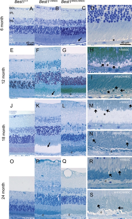
Figure 4.
Development and progression of serous retinal detachments and debris-filled apical deposits in Best1W93C knock-in mice. Representative micrographs of Toluidine blue-stained thick sections from mice aged 6 (A–D), 12 (E–I), 18 (J–N) or 24 (O–S) months of age as indicated in the left margin. Genotypes for A–C, E–G, J–L and O–Q are indicated at the top of the figure. Panels D, H, I, M, N, R and S are identical to panels C, F, G, K, L, P and Q, respectively, but were photographed at a higher magnification. Their genotypes are indicated in the upper right hand corner of the panel. Serous detachments (arrows in C, F and K, asterisks in D, H and M) were noted as early as 6 months of age. Following detachment, we often found piles of RPE microvilli laying horizontal and containing debris (d) including OS. Disruption of OS was noted in many animals as early as 12 months of age (arrows in H, I, M, N, R and S).

