Fig. 6.
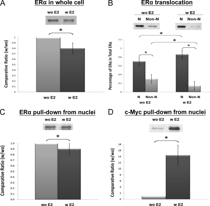
A, Western blotting of ERα in whole cell lysate with (w) and without (w) a 24-h E2 (10−8 m) treatment (n = 4). B, translocation of ERα from the non-nuclei (non-N) to the nuclei (N) of MCF-7 cells upon 24-h stimulation with E2 (10−8 m) (n = 8). C, ERα in the AuNP-ERE pulldown from the nuclear fractions with and without E2 treatment (n = 5). D, c-Myc in the AuNP-ERE pulldown from the nuclear fractions with and without E2 treatment (n = 3). Significant differences (p < 0.05) are indicated with the star (*).

