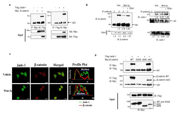
Figure 1.
Jade-1 and β-catenin interact. (a) In vivo interaction of Jade-1 and β-catenin. Extracts (600 μg protein) from transiently transfected 293T cells were immunoprecipitated (IP) with 1 μg monoclonal Myc-tag or Flag-tag antibodies. Co-immunoprecipitated β-catenin or Jade-1 was detected by immunoblotting. Whole cell lysates (WCL) (10%) were probed for input. Representative immunoblot of 4 experiments. (b) The interaction of endogenous Jade-1 and endogenous β-catenin is increased in Wnt-off phase. IPs were performed with WCL (500 μg protein) of 293T cells pretreated with vehicle (PBS + 0.1% bovine serum albumin-BSA) or 50 ng Wnt-3a ligand in PBS + 0.1% BSA, with or without 50 ng DKK1, using 1 μg of either rabbit polyclonal Jade-1 antibody (J1) or pre-immune rabbit serum (C). The co-immunoprecipitated β-catenin was detected by immunoblot with monoclonal β-catenin antibody. β-catenin was immunoprecipitated as described above using monoclonal β-catenin antibody (β-cat) and isotype control (C). Jade-1 was detected by immunoblot using Jade-1 antiserum. WCL (10%) were probed for input. Densitometry was performed to quantitate β-catenin and Jade-1. The amount of Jade-1 and β-catenin immunoprecipitated was normalized using IgG. Representative immunoblot of 3 experiments. (c) Co-localization of endogenous Jade-1 and endogenous β-catenin is increased in Wnt-off status. The 293T cells pretreated with vehicle or Wnt-3a (200 ng) for 4 h were fixed and incubated with monoclonal β-catenin and polyclonal Jade-1 antibodies followed by Alexa 594 donkey anti-mouse and Alexa 488 goat anti-rabbit as secondary antibodies. Profile plots were generated using NIH ImageJ to demonstrate quantitative Jade-1 and β-catenin protein distribution. The profile plot represents the signal intensity of each fluorophore along a single line across the midpoint of a representative cell. The X axis represents the distance in pixels through the length of a single cell, and the intensity of each fluorophore is plotted on the Y axis. A representative image from 4 experiments is shown. Scale bar = 10 μm. (d) Identification of the domain of β-catenin required for Jade-1 binding. Extracts from transiently transfected 293T cells were immunoprecipitated. WCL (10%) were probed for input. β-catenin delC and delN constructs lack the C terminus and N terminus of the protein, respectively (Supplementary Fig. S1a). β-catenin S33A is a naturally occurring, cancer-causing amino acid substitution mutant. Representative immunoblot of 3 experiments.

