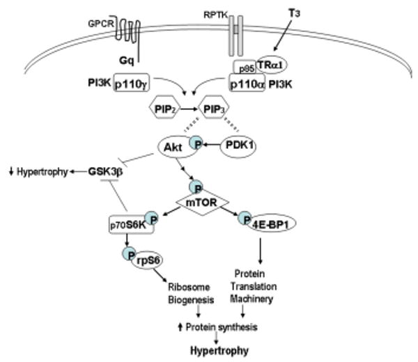
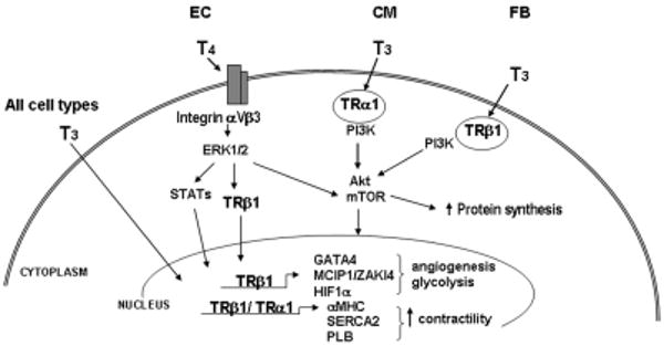
Figure. Schematic representation of thyroid hormone signaling in cardiac hypertrophy.


(A) In physiological hypertrophy, T3 binding to cytoplasmic TRα1 or growth factor binding to receptor protein tyrosine kinases (RPTK) activates PI3K isoform p110α through direct interactions with the p85 subunit of PI3K. p110α phosphorylates phosphatidylinositol PIP2 at the 3′ position to produce PIP3, which serves to bind phosphatidylinositol-dependent kinase (PDK1) and Akt, allowing PDK1 to phosphorylate Akt. Activation of Akt then leads to activation of mTOR (mammalian target of rapamycin) which regulates protein synthesis through downstream targets including S6 kinase (p70S6K) and initiation factor eIF4E binding protein (4E-BP1), resulting in increased rates of protein translation and ribosome biosynthesis, leading to cardiomyocyte hypertrophy. Akt and S6K also phosphorylate and inhibit glycogen synthase kinase (GSK3β), an inhibitor of protein synthesis, thereby promoting cardiac hypertrophy. In contrast, pathological hypertrophic agonists (i.e., angiotensin, catecholamines) acting through G-protein coupled receptors (GPCR) and Gq, activate the Akt/mTOR pathway by associating with the p110γ isoform of PI3K.
(B) Reported effects of thyroid hormones (T3, L-tri-iodothyronine; T4, L-thyroxine) on various cell types including endothelial cells (EC), cardiomyocytes (CM) and fibroblasts (FB). T3 activation of the PI3K/Akt/mTOR signal transduction pathway as been shown to be initiated by binding to cytoplasmic thyroid hormone receptors (TRα1 and TRβ1), resulting in increased protein synthesis and activation of a hypertrophic gene program. In endothelial cells, T4 has been reported to bind to a plasma membrane integrin receptor (αVβ3) and to activate the mitogen-activated protein kinase (ERK1/2) signaling pathway, leading to TRβ1 phosphorylation and/or translocation to the nucleus where its transcriptional activity is enhanced. Furthermore, T3 can enter the nucleus directly in virtually all cells and bind to nuclear TRs that regulate expression of target genes that increase cardiac contractility, stimulate angiogenesis, regulate cell metabolism and provide cytoprotection. (MCIP, inhibitor of calcineurin; HIF, hypoxia-inducible factor; MHC, myosin heavy chain; PLB, phospholamban; SERCA2, sarcoplasmic reticulum calcium ATPase; GATA4, transcription factor).
