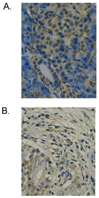
Figure 5. Immunohistochemistry for MnSOD.
Loss of MnSOD expression in pancreatic ductal cells from pancreatic resections of adenocarcinoma is shown. Immunohistochemistry for MnSOD expression using the avidin-biotin peroxidase complex method was performed on pancreatic specimens previously fixed in formalin and embedded in paraffin. A quantitative digital imaging methodology was used to examine MnSOD staining in the pancreatic tissue. Cytoplasmic regions of pancreatic ductal cells were identified and digitized. Mean gray-level pixel values were then obtained (A). Strong staining is seen in the cytoplasm in cells from normal pancreas. Staining is nearly undetectable in cells from pancreatic cancer resections with a marked decrease in the mean gray level value when compared to normal pancreas (B). All experiments using animals or human samples should be reviewed and approved by the Institutional animal care and use committee. Bar = 50 μm.

