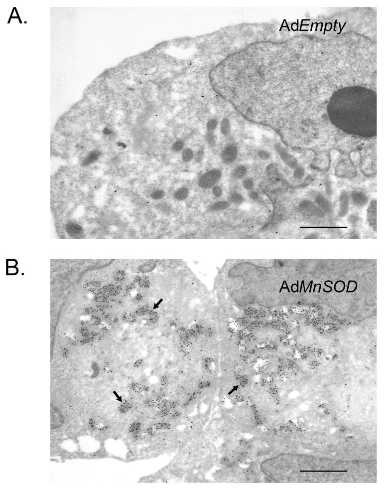
Figure 6. Sub-cellular localization of MnSOD by immunogold.
MIA PaCa-2 human pancreatic cancer cells were infected with adenoviral vectors containing the cDNA for MnSOD. Ultrastructural examination was performed by Dr. Terry Oberley at the University of Wisconsin. Sections were treated with primary MnSOD antibody overnight, washed, and treated with gold-conjugated goat anti-rabbit immunoglobulin, fixed and stained. Labeling of MnSOD was extremely light in cells treated with the AdEmpty vector (A). MIA PaCa-2 cells treated with AdMnSOD demonstrated increases in labeling (arrow) in the mitochondria (bottom panel) (B). All experiments using animals or human samples should be reviewed and approved by the Institutional animal care and use committee. Bar = 5 μm

