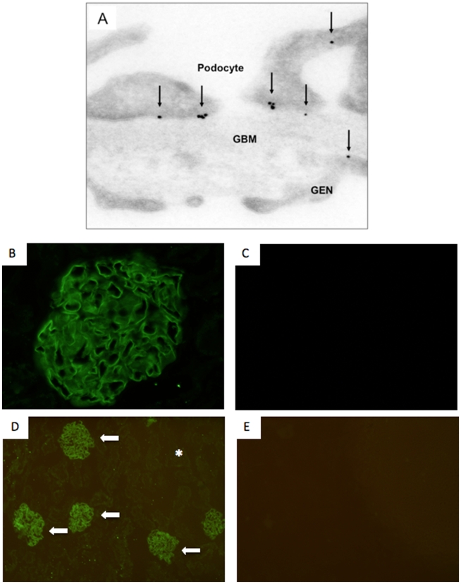
Figure 2. Affinity of the anti-podocyte antibody.
The antibody binds to the apical, basal and the slit diaphragm regions of the podocyte (A). A small number of immunogold particles were present in the glomerular basement membrane (GBM) and endothelial cells (GEN). The arrows indicate examples of immunogold particles. Sheep anti mouse podocyte antibody staining was detected only in the glomerulus (B + D arrows) and was absent in the tubular compartment (D asterix). Control animals injected with normal sheep IgG showed no staining (C + E).

