
Figure 1.
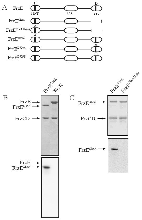
FrzECheA in vitro autophosphorylation. (A) A schematic of the domain structure of FrzE and mutants constructed. HPT refers to the histidine phospho-transfer domain and CA, refers to the catalytic domain. The conserved residues H-49 and D-709 are indicated. (B) A Coomassie stained gel above and a phosphorimage below of an SDS-PAGE gel containing FrzCD, FrzA, FrzECheA (lane1), FrzE (lane 2), and [γ-32P]-ATP. (C) A Coomassie stained gel above and a phosphorimage below of an SDS-PAGE gel containing FrzCD, FrzA, FrzECheA (lane1), FrzECheA H49A (lane2), and [γ-32P]-ATP. Note: FrzA is a small protein that has migrated off all gels in (B) and (C).