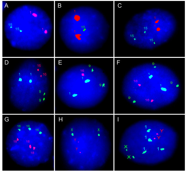
Figure 1.
Two- and three-color I-FISH with centromeric DNA probes. (A) normal diploid nucleus with two signals for chromosome 1 and chromosome 15; (B) monosomic nucleus with two signals for chromosome 1 and one signal for chromosome 15; (C) trisomic nucleus with two signals for chromosome 1 and three signals for chromosome 15; (D) normal diploid nucleus with two signals for chromosome 1, chromosome 9 and chromosome 16; (E) monosomic nucleus with two signals for chromosome 1 and chrosmome 9 and one signal for chromosome 16; (F) trisomic nucleus with two signals for chromosome 1 and chromosome 16 and three signals for chromosome 9; (G) triploid nucleus with three signals for chromosome 16 and chromosome 18; (H) tetraploid nucleus with two signals for chromosome X and chromosome Y; (I) tetraploid nucleus with two signals for chromosome X and chromosome Y, and four signals for chromosome 1.

