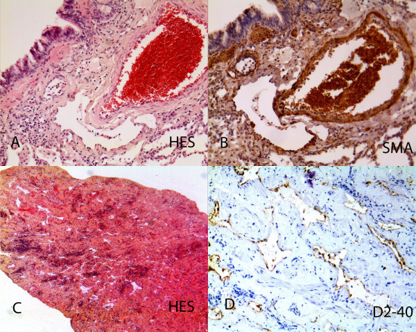
Figure 6.
Pathological findings: Lung locations. A: Peribronchiolar dilated lymphatics (HES × 400). B: Foci of smooth muscle actin positivity alongside dilated lymphatic (SMA × 200). C: Parietal biopsy at low magnification showing its thickening, with numerous lymphatic cavities and inflammation (HES × 100). D: Higher magnification of the D2-40 lining of vascular cavities with some smooth muscle bundles in between (SMA × 200).

