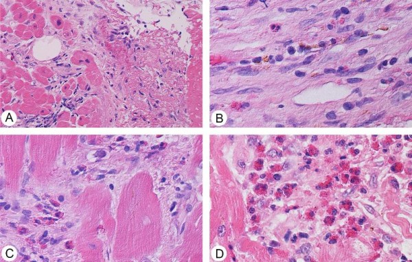
Figure 5.

Endomyocardial biopsy showing the following: (A) Organizing thrombus in small vessels of endocardium (Hematoxylin and Eosin staining, ×20 magnification). (B) Older areas show organized endocardial scar with rare eosinophils and hemosiderin-laden macrophages (Hematoxylin and Eosin staining, ×40 magnification). (C) Close-up of intact and degranulating eosinophils in the interstitial space, without myocyte necrosis (Hematoxylin and Eosin staining, ×40 magnification). (D) A larger cluster of non-degranulated eosinophils (Hematoxylin and Eosin staining, ×40 magnification).
