Figure 1.
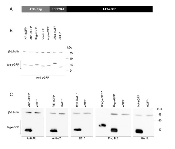
SDS-PAGE analysis of tag-eGFP fusion proteins in LV transduced HEK293T cells. A) Schematic representation of the tag-eGFP constructs. Peptide sequences are listed in table 1. B) Overexpressed proteins were detected with an in-house anti-eGFP antibody. C) The same lysates as in B) were analyzed with the different anti-tag antibodies. 2 μg of cell lysate was loaded and antibody dilutions were as follows: anti-eGFP (1/10000), anti-AU-1 (1/25000), anti-V5 (1/250000), 9E10 (1/10000), FlagM2 (1/30000) and HA 11 (1/50000). β-tubulin was used as loading control. * cell lysate of 3flag-eGFP was loaded at 4 ng to avoid saturation of the western blot signal.

