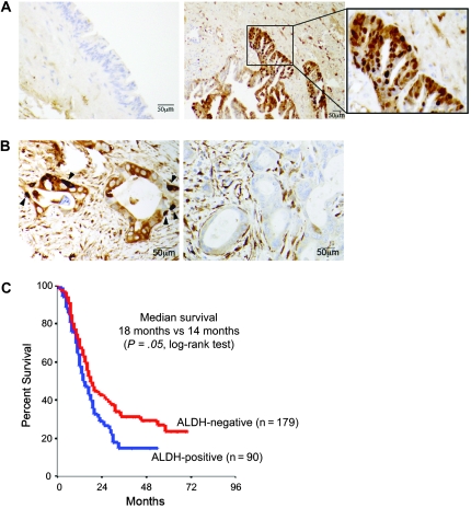
Figure 1.
Association between aldehyde dehydrogenase (ALDH) expression in primary pancreatic tumors and patient survival. A) Immunohistochemical localization of ALDH protein expression in pancreatic intraepithelial neoplastic lesions. Representative images show staining for ALDH (brown) in a high-grade pancreatic intraepithelial neoplastic lesion (ie, PanIN-3 or carcinoma in situ; right panel and inset) and in a low-grade pancreatic intraepithelial neoplastic lesion (ie, PanIN-2; left panel). B) Immunohistochemical staining of ALDH in primary human pancreatic adenocarcinoma. Left panel shows an ALDH-positive tumor in which intensely staining cells are found mostly at the base of cancerous glands (arrowheads). Right panel shows an ALDH-negative tumor. C) Kaplan–Meier analysis of overall survival for patients who underwent pancreatectomy based on tumor ALDH expression by immunohistochemistry. At 24 months, the number of patients at risk with ALDH-positive and ALDH-negative tumors was 27 (proportion surviving = 0.30, 95% confidence interval [CI] = 0.22 to 0.42) and 67 (proportion surviving = 0.39, 95% CI = 0.32 to 0.47), respectively. At 48 months, the number of patients at risk with ALDH-positive and ALDH-negative tumors was 9 (proportion surviving = 0.15, 95% CI = 0.09 to 0.25) and 33 (proportion surviving = 0.23, 95% CI = 0.18 to 0.31), respectively. At 72 months, the number of patients at risk with ALDH-positive and ALDH-negative tumors was 2 (proportion surviving = 0.11, 95% CI = 0.05 to 0.21) and 16 (proportion surviving = 0.17, 95% CI = 0.12 to 0.24).

