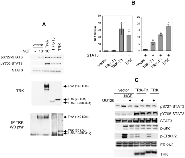
Figure 1. STAT3 activation by TRK oncogenes.
(A) Western blot analysis of PC12 cells transfected with empty pRC/CMV vector, TRKA, TRK-T3, and TRK cDNAs. NGF treatment (50ng/ml, 10′) is indicated. Cell lysates and immunocomplexes were separated by SDS PAGE as described in Material and Methods and immunoblotted with indicated antibodies. In the bottom panel two distinct exposures of the same blot were used for documenting TRKA or TRK oncoproteins phosphorylation. (B) HeLa cells were co-transfected with pM67 and pRL-TK in combination with the indicated TRK oncogene cDNAs in the absence (left graph) or presence (right graph) of STAT3 cDNA and assayed for STAT3-dependent luciferase activity 48 hours later. Activity is expressed as the ratio of luciferase/renilla activity, and reported as fold-inductions over empty vector (left panel) or STAT3 (right panel) (RLA: Relative Luciferase Activity). The data represent the mean values ± SD of triplicate samples. Similar results were obtained in three independent experiments. (C) MAPK involvement in TRK-induced Stat3 phosphorylation. Western blot analysis of PC12 cells transfected with empty pRC/CMV vector, TRK-T3 or TRK cDNAs. NGF (10′, 50ng/ml) and UO126 (16 hr, 10 µM) treatments are indicated. Immunoblotting was performed with the indicated antibodies.

