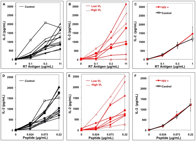
Figure 3. Antigen presentation by HLA-DR1+ MN from individual HIV-infected and uninfected subjects.
In all four panels, MN (5×10⁴/well) were incubated with soluble antigen or peptide and T cell hybridoma (1×10⁵/well) for 24 hrs. Viremic HIV-infected individuals with VL>1000 are High VL and VL<1000 are Low VL. A. Control MN and soluble RT, B. HIV+ MN and soluble RT, C. Combined data on soluble RT presentation, D. Control MN and peptide, E. HIV+ MN and peptide, F. Combined data on peptide presentation.

