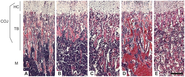
Figure 1. Collagen X Tg and KO mice have an altered chondro-osseous junction.
Hematoxylin and eosin staining of longitudinal sections of tibia from (A) week-3 C57Bl/6 wild type (WT), (B) C57Bl/6 congenic collagen X transgenic (Tg), (C) null (KO), and perinatal lethal (D) Tg, and (E) KO. The chondro-osseous junction (COJ) is shown, including hypertrophic cartilage (HC), trabecular bone (TB), and marrow (M). Note diminished trabecular bony spicules in all collagen X Tg and KO mice, with the greatest reductions in (D) and (E), with a concomitant increase in erythrocytes in the marrow. Bar = 150 µm.

