Figure 8.
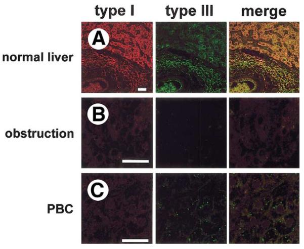
Expression of the type I InsP3R in normal and cholestatic human liver. Confocal immunofluorescence images were obtained from paraffin-embedded liver biopsy specimens. Each tissue section was double-labeled with antibodies directed against the type I InsP3R (red) and type III InsP3R (green). Images include both hepatocytes and cholangiocytes so that relative InsP3R expression in the 2 cell types can be compared. (A) Normal liver. Expression of the type I InsP3R is low in cholangiocytes relative to hepatocytes, whereas the type III InsP3R is localized to the apical region of cholangiocytes and is absent in hepatocytes. Note that the type I InsP3R is distributed diffusely in hepatocytes, as has been reported.67. (B) Bile duct obstruction. Expression of type I InsP3R is preserved in hepatocytes, but neither type I nor type III InsP3R is detectable in cholangiocytes. Findings are representative of 3 separate biopsy specimens. (C) Primary biliary cirrhosis. Expression of type I InsP3Rs is preserved in hepatocytes, but neither type I nor type III InsP3Rs are detected in cholangiocytes. Findings are representative of 3 separate biopsy specimens. Each scale bar = 30 μm.

