
Figure 3.
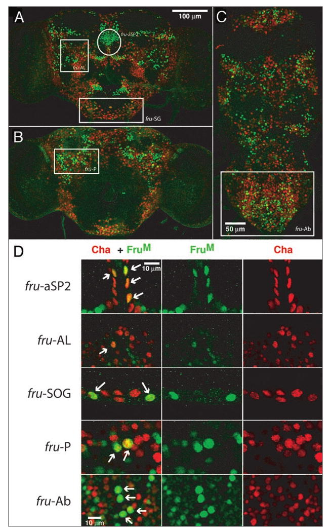
Co-localization of FruM and cha-Gal4 driven positive neurons. Anterior (A) and posterior (B) brain and ventral nerve cord (C) of cha-GAL4/UAS-RedStinger males stained with FruM (green). (D) Higher magnification of selected clusters highlighted in (A–C) (aSP2-anterior region of the superior protocerebrum; SG-subesophageal ganglion; AL-broadly scattered cells above the antennal lobe; P-broadly scattered in posterior region, ventral to calyx; Ab-abdominal ganglion region). Arrows show examples of neurons showing co-localization of FruM and cha-Gal4 labeling. See Table S1 for complete counts.