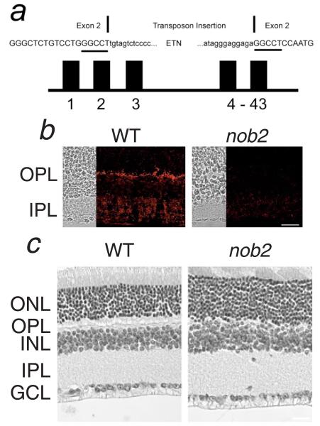
Fig. 1.
(a) Diagram of Cacna1f, indicating the location of the intragenic insertion into exon 2. Exon 2 nucleotides are shown as capitals. The 6-bp duplication at the insertion site is underlined. (b) Immunofluorescent staining for α1F is localized to the OPL and the IPL of the WT retina but is absent in the nob2 retina. (c) Comparison of retinal anatomy in adult WT and nob2 mice. In the nob2 retina, the OPL is disorganized but other retinal layers appear normal. GCL, ganglion cell layer; IPL, inner plexiform layer INL, inner nuclear layer; OPL, outer plexiform layer; and ONL, outer nuclear layer. Scale bar indicates 20 μm (b) or 40 μm (c).

