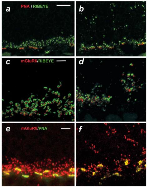
Fig. 5.
Synaptic abnormalities in nob2 retina. (a–f) micrographs of vertical sections through WT (a, c, & e) or nob2 (b, d, & f) retinas immunolabeled with PNA (red)/Ribeye (green; [a & b]), mGluR6 (red)/Ribeye (green; [c & d] or mGluR6 (red)/PNA (green; [e & f]. Scale bar indicates 10 μm (a, b) or 5 μm (c–f). Mice were studied at 5 months of age.

