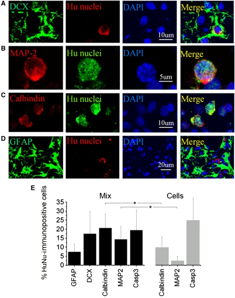
Figure 3.
Cell type-specific marker expression in transplanted NPCs in vivo. The brains were removed 8 weeks after transplantation of NPC/Matrigel cultures, sectioned through the frontal cortex adjacent to the lesion cavity, and stained with antibodies against cell-type markers (left panels) and HuNu (panels second from left). Nuclei were counterstained with DAPI (blue; panels second from right). HuNu-immunopositive cells expressed neuronal markers, including (A) DCX (green), (B) MAP-2 (red) and (C) calbindin (red), but not (D) the astroglial marker GFAP (green). (E) Cell counts in the periinfarct cortex after transplantation of human ESC-derived NPCs alone (Cells) or NPC/Matrigel cultures (Mix) were expressed as a percentage of all HuNu-immunopositive cells (mean±s.e.m., n=3 rats per condition). *P<0.05.

