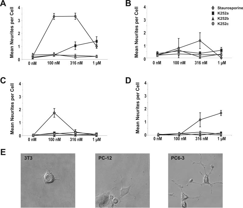
Figure 2. Effects of staurosporine and related compounds on neuronal and non-neuronal differentiation.
Treatment of RGC-5 (A), 3T3 (B), PC-12 (C), and PC6-3 (D) cells with staurosporine caused significant levels of differentiation. K252b and K252c had no effect on neurite extension. Treatment of RGC-5 cells with K252a caused significant primary neurites to extend (1.0 ± 0.3 at 316 nM vs. 0.17 ± 0.07 at 0 nM; p < 0.001), but RGC-5 cells treated with K252a extended fewer neurites than cells treated with staurosporine (1.0 ± 0.3vs. 3.3 ± 0.2 respectively at 316 nM; p < 0.001). Similarly, process formation was seen with staurosporine, but not K252a, K252b, or K252c in 3T3, PC-12, and PC6-3 cells. Photomicrographs (E) illustrate process extension of 3T3 cells treated with 316 nM SS, PC-12 cells treated with 316 nM SS, and PC6-3 cells treated with 1 μM SS.

