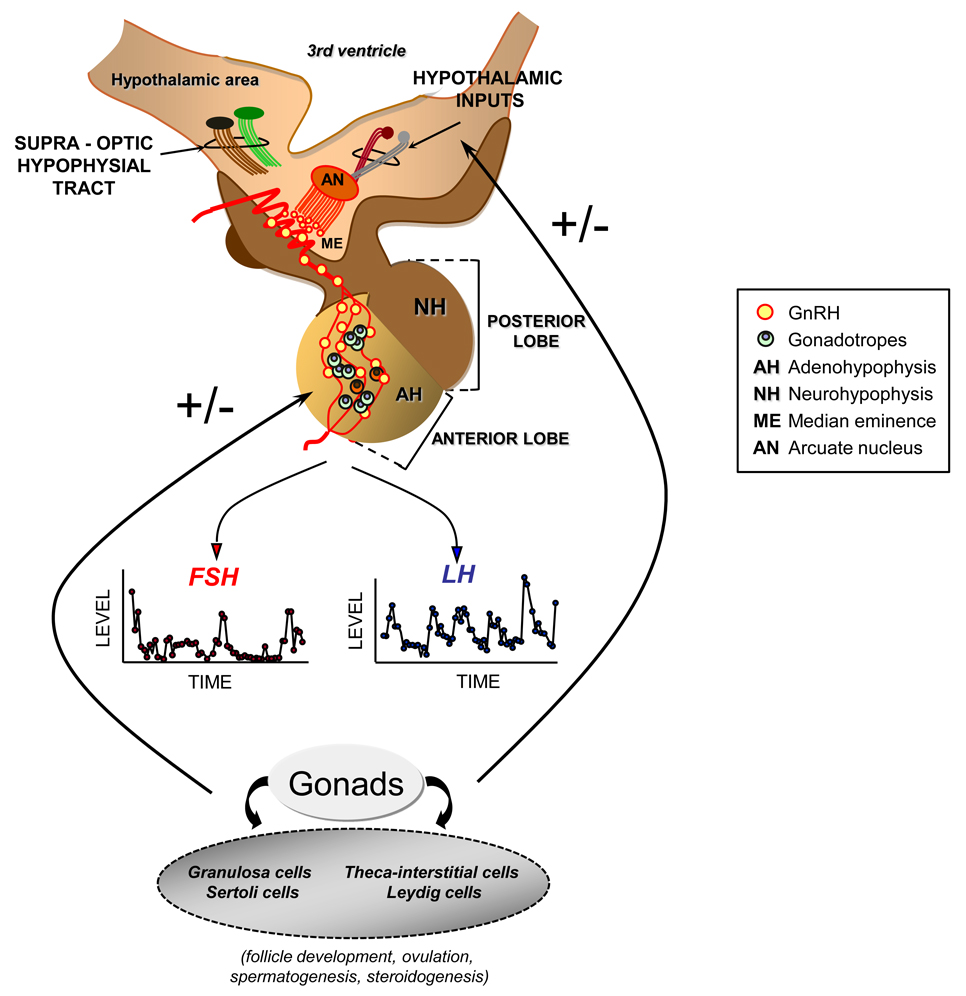
Figure 1.
Functional relations of the hypothalamic–pituitary axis. Gonadotropin-releasing hormone (GnRH) is synthesized and secreted by specialized neurons located mainly in the arcuate nucleus (AN) of the medial basal hypothalamus and the preoptic area of the anterior hypothalamus. GnRH producing neurons project to the median eminence (ME) where they terminate in an extensive plexus of boutons on the primary portal vessel, which delivers GnRH to its target cell, the gonadotrope of the adenohypophysis (AH). The secretion and interaction of GnRH with its cognate receptor occurs in a pulsatile and intermittent manner; such episodic signaling allows the occurrence of distinct rates and patterns of synthesis and pulsatile release of luteinizing hormone (LH) and follicle-stimulating hormone (FSH). These gonadotrophic hormones are responsible for stimulating the synthesis and secretion of gonadal hormones and for affecting the process of gametogenesis. The characteristics of the pulsatile release of GnRH, LH, and FSH appear to be positively or negatively regulated by several hypothalamic neurotransmitters (e.g., adrenergic and opioidergic regulation), as well as by the gonadal hormone environment.

