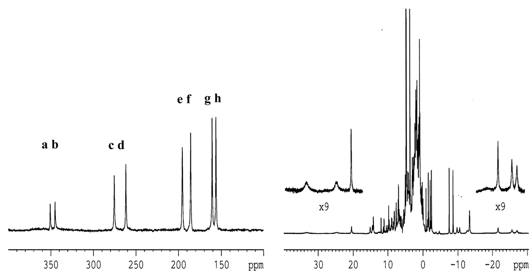
FIGURE 1.
The 1D 1H NMR spectrum of DvRd(Ni) at 302K. The low field contact shifted Hβ protons from the 4 binding cysteines can be observed between 350 and 150 ppm (a–h). Other contact and pseudocontact shifted peaks can be seen outside of the diamagnetic envelope up to +30 and down to −30 ppm. The difference in peak intensity seen for the low field shifted peaks is due to an uneven excitation profile.

