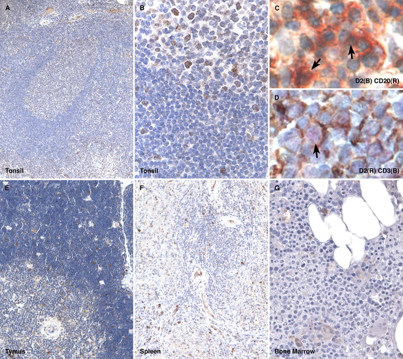
Figure 1. Cyclin D2 expression in normal hematopoietic tissue.
Cyclin D2 staining is found in a subset of cells in the germinal center, mantle and marginal zones and in the paracortex of a normal tonsil (A and B); double immunohistochemical labeling for cyclin D2 (brown) and CD20 (red) show co-localization of staining in a subset of B-cell (arrows, C); double immunohistochemical labeling for cyclin D2 (red) and CD3 (brown) show co-localization of staining in a subset of T-cell (arrow, D); scattered cells in the cortex and medulla of the normal thymus show cyclin D2-positive cells (E); the normal spleen shows staining in scattered lymphoid cells in the red and white pulp and in endothelial cells but is lacking in splenic marginal zones (F); the normal bone marrow shows staining in scattered lymphoid and plasma cells but is not found in a significant proportion of erythroid or myeloid precursors or megakaryocytes (G).

