
Fig. 1.
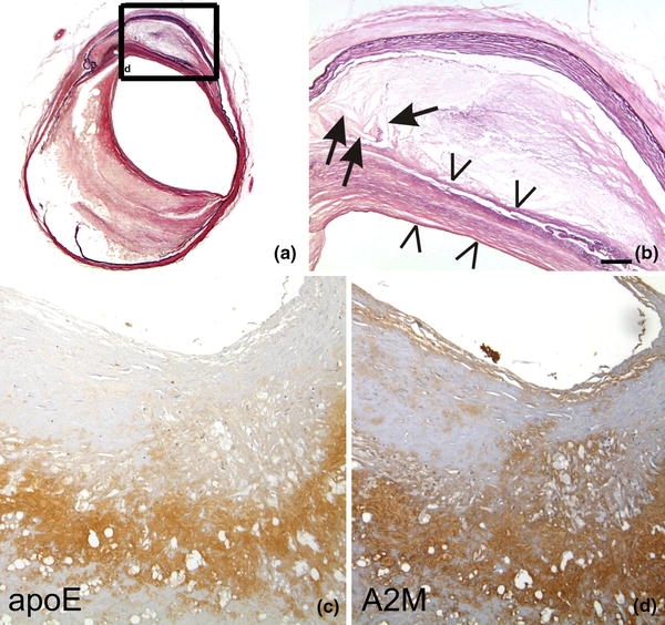
a, b The left internal carotid artery of a 79-year-old man exhibits severe atherosclerotic changes (Type 5 according to Stary [119]). There is a thinning of the lamina media, proliferation and lipid accumulation in the intima including cholesterol clefts (arrows). The necrotic core is covered by a fibromuscular tissue layer (arrowheads) indicative for Stary Type 5 lesions. b Corresponds to a high magnification view of the boxed area in (a). ApoE (c) and A2M (d) occur in the plaque core of an AS plaque. Staining in (a, b) Elastica van Gieson (EVG), c anti-apoE [Covance (Dedham, USA), D6E10, 1/500, formic acid and microwave pretreatment], d anti-A2M [BioMac (Germany, Leipzig), polyclonal rabbit, 1/5,000]. The calibration bar in (b) corresponds to: a 400 μm, b 90 μm, c, d 70 μm