
Fig. 2.
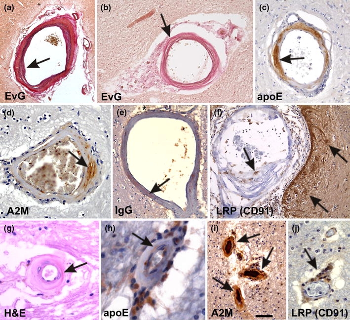
Small vessel disease-related changes. a A leptomeningeal artery shows intima proliferation and a splitting of the internal elastic lamina (arrow). These changes are related to small vessel arteriosclerosis/atherosclerosis. b A white matter artery exhibits fibrosis, lipohyalinosis of the vessel wall, and fibrinoid necrosis (arrow). Lipohyalinosis affected vessels exhibit the plasma proteins apoE (c), A2M (d), and IgG (e) within the vessel wall (arrows in c–e) indicating the leakage of plasma proteins into the vessel wall and into the perivascular space (asterisk in e). f Macrophages within the lipohyalinotic lesions and perivascular astrocytes strongly exhibit the A2M and apoE receptor LRP (CD91) (arrows) indicating that these cells are capable of taking up A2M and apoE. g Arteriolosclerosis of a white matter artery shows severe hyalinization (arrow) of the vessel wall. h–j ApoE and A2M were observed within the vessel wall of arteriolosclerotic vessels (arrows in h, i). Within the enlarged perivascular spaces high numbers of apoE (h), A2M (i), and LRP-positive cells (arrow in j) were observed indicating that these perivascular cells accumulate apoE and A2M due to an insufficient perivascular drainage. These perivascular macrophages are often Prussian blue negative and do not necessarily represent hemorrhagic residues [129]. Stainings in a–j as indicated. Anti-apoE and anti-A2M staining was performed as indicated in Fig. 1. For anti-IgG and anti-LRP immunohistochemistry the following antibodies were used [anti-IgG: polyclonal goat; Biomeda, Foster City, CA; 1/100; microwave pretreatment; anti-LRP (anti-CD91): α2-M-R-II2C7; BioMac, Leipzig, Germany; 1/150; microwave and protease pretreatment]. The calibration bar in i corresponds to: a 300 μm, b 80 μm, c 40 μm, d, j 35 μm, e, f 60 μm, g 20 μm, h 16 μm, i 50 μm. a and b are reproduced from Thal et al. 2003 [129] with kind permission