
Fig. 3.
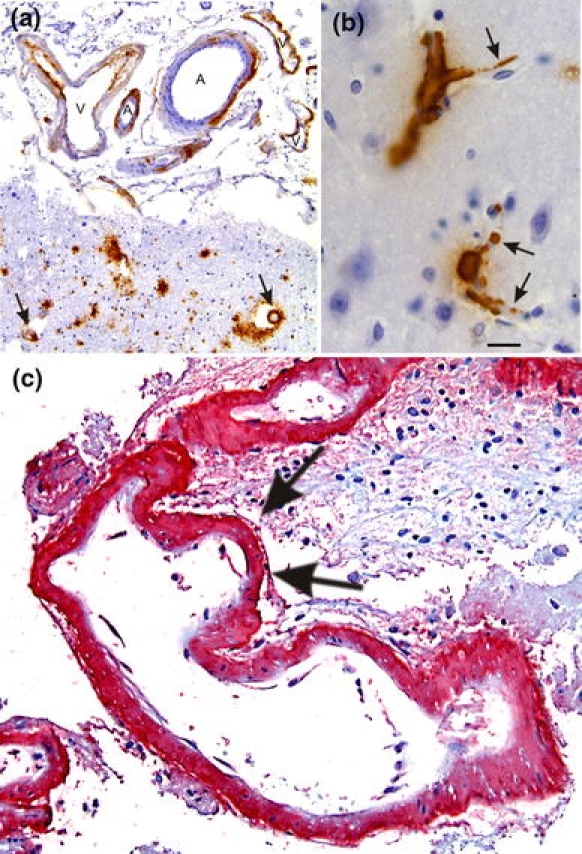
Cerebral amyloid angiopathy (CAA). a Aβ deposition in the vessel wall of leptomeningeal arteries (A) and veins (V) as well as in cortical arteries (arrows). b Capillary CAA is characterized by Aβ deposits at the basement membrane of cortical capillaries (arrows). c Severe CAA in a case of CAA-related hemorrhage. The CAA-affected artery exhibits multiple aneurysmal dilations of the vessel wall as indicated by arrows. Aβ deposits are stained in red (permanent red; DAKO, Glostrup, Denmark) with an antibody against Aβ17–24 (4G8, Covance, Dedham, USA, 1/5,000, pretreatment with formic acid). The same antibody was also used in figures a and b but 3,3-diaminobencidine–HCl was used as chromogen. The calibration bar in b corresponds to: a 85 μm, b, c 20 μm