
Fig. 4.
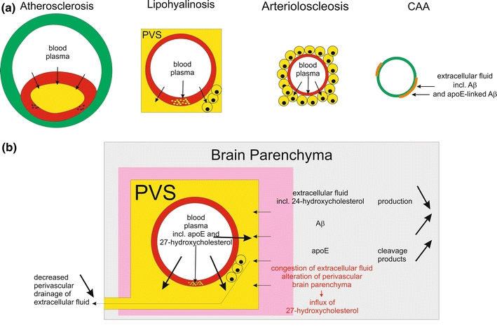
Plasma protein leakage induced by vessel disorders and its relation to perivascular alterations of the brain parenchyma. a This schematic representation shows that plasma proteins occur (1) in the plaque cores of AS plaques, (2) in the vessel wall of lipohyalinotic vessels as well as in the perivascular space and in macrophages within the perivascular space, and (3) in the vessel wall of arteriolosclerotic vessels as well as in accompanying macrophages. CAA, on the other hand, is characterized by the deposition of proteins of the extracellular fluid of the brain, i.e. Aβ [15] and apoE [133]. b Impact of plasma protein leakage into the brain. Physiologically, extracellular fluid is drained into the perivascular space and along the vascular basement membranes [16, 60, 155]. In the event of SVD, there is plasma protein leakage into the vessel wall and into the perivascular space [139] resulting in (1) a competition between leaking plasma and extracellular fluid from the brain for perivascular drainage and (2) the congestion of extracellular fluid leading to the accumulation and/or alternative processing of proteins of the extracellular fluid, and 3) the influx of the peripheral cholesterol metabolite 27-hydroxycholesterol into the brain [46, 127, 139]. The influx of 27-hydroxycholesterol into the brain is accompanied by decreased levels of brain derived 24-hydroxycholesterol indicating a reduction in the cerebral 24-hydroxycholesterol production [46]