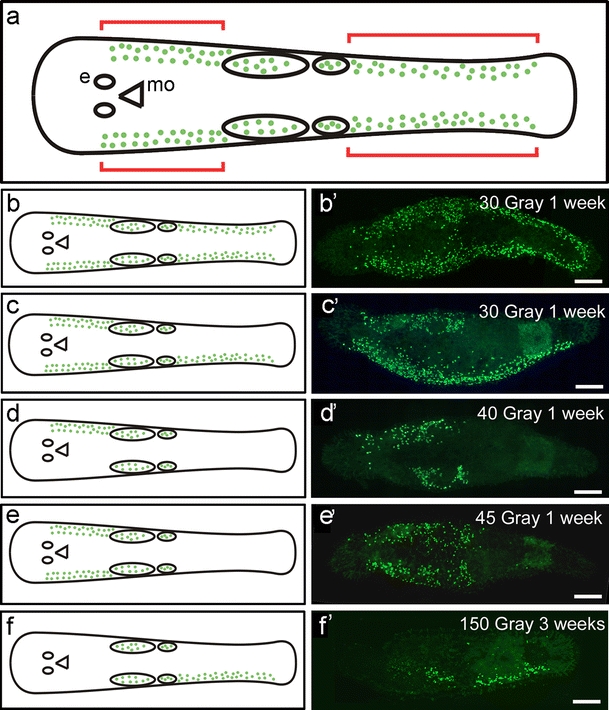
Fig. 8.

Stem cell recovery in lateral compartments. a Representation indicating the recovery of somatic stem cells (e eye, mo mouth opening, green dots BrdU-labelled cells, anterior ovals testes, posterior ovals ovaries, red brackets region in which stem cell recovery was followed). b-f Schematic drawings of the recovery of cell proliferation (BrdU). b’–f’ Expression of macpiwi expression. b, b’ Recovery occurred in all regions. c-f’ Recovery of proliferating cells in different regions of the animal. Bars 100 µm
