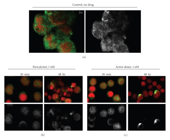
Figure 7.
Localization of VDR in pig parathyroid cells. Cells were incubated without (a) or with 1 nM paricalcitol (b) or active doxercalciferol (c) for 30 minutes or 48 hours, and then stained for VDR (green) and nucleus (red). The VDR staining alone was also shown in black and white. (a): 3× magnification; (b) and (c): 2× magnification; doxer: doxercalciferol.

