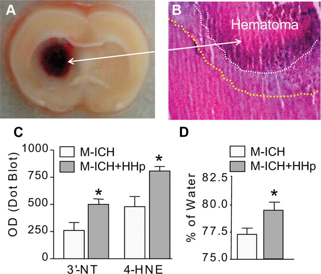
Figure 1.
HHp aggravates ICH-mediated damage. A, Photograph of representative rat brain coronal section illustrating location of hematoma produced with intrastriatal injection of lysed blood (M-ICH). B, Representative photomicrograph of H&E-stained cryosection showing edema surrounding hematoma (outlined with dotted lines), as captured at 24 h after M-ICH. C, Bar graph illustrating the densitometrical analysis of immunoreactivity (immuno-dot blots technique) for 3′-NT and 4-HNE in the hematoma-affected tissue homogenates of normohaptoglobinemic control (ICH) and HHp (ICH+HHp) rats 24 h after M-ICH. The data are expressed as mean ± SEM (n = 5). *p ≤ 0.05 from the control (ICH). D, Bar graph illustrating brain edema measured by wet-to-dry weight ratio (percentage of water) at 24 h after M-ICH in the control (ICH) and HHp (ICH+HHp) rats. The data are expressed as mean ± SEM (n = 7). *p ≤ 0.05 from the control (ICH).

