Figure 6.
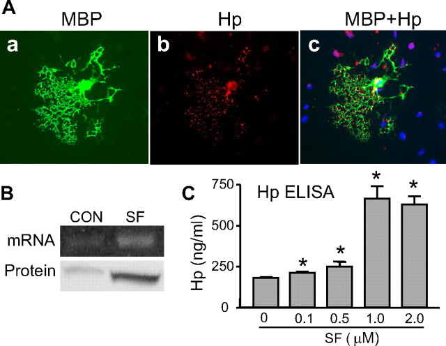
Hp is expressed by oligodendroglia and its expression is stimulated by SF. A, Photomicrograph of MBP-immunolabeled oligodendrocyte expressing Hp. The mixed neuronal–glial cocultures were incubated with 1 μm SF for 24 h and then immunolabeled for MBP (a) (green) and Hp (b) (red). c, Merged image of MBP and Hp. The nuclei were visualized with Hoechst 33258 (blue). Note that, among all cells in the field, only MBP-positive cell is immunopositive for Hp. Hp proteins are detected in the soma and fine processes of MBP-positive cells. Scale bar, 100 μm. B, Photograph of RT-PCR product for Hp on representative agarose gel (mRNA, top panel) and Hp immunoblot (protein, bottom panel), illustrating levels of Hp expression in the oligodendroglia-enriched cultures in absence (CON) or presence of SF (1 μm for 24 h). C, Bar graph quantifying Hp protein in the oligodendrocyte-enriched culture media, at 48 h after treatment with 0.1∼2 μm SF, determined by ELISA. *p < 0.05 from vehicle control.

