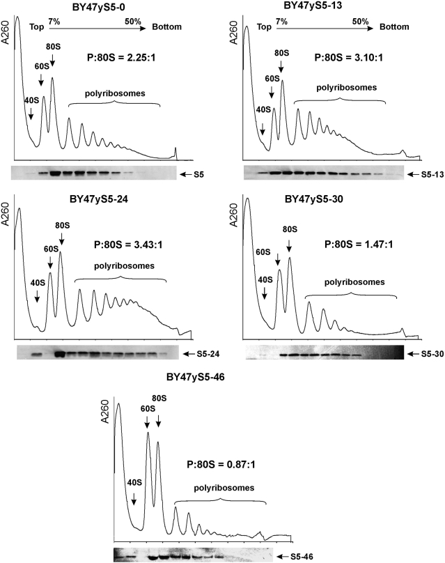
Figure 3.
Ribosome profiles of the wt and mutant yeast strains containing truncated versions of the rpS5 protein and rpS5 western blot analyses. Extracts from isogenic wt (BY47yS5-0) and rpS5 mutant (BY47yS5-13, BY47yS5-24, BY47yS5-30 and BY47yS5-46) strains were resolved by velocity sedimentation on 7–50% sucrose gradients. Fractions were collected while scanning at A254. The positions of different ribosomal species are indicated. The ratio of the area under the polysomal (P) and 80S peaks is shown (P:80S). The data recorded by the PeakTrak program (ISCO gradient density gradient fractionation system) were exported to ASCII format and analyzed by UVProbe 2.10 Shimadzu software to assess the 80S and polysomal peak areas. Western blot analysis of individual fractions was done using an antibody directed against the AIKKKDELERVAKSNR C-terminally conserved rpS5 peptide.

