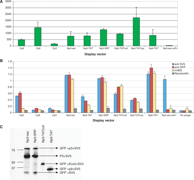
Figure 3.
Expression and display of GFP using different display vectors. (A) The fluorescence levels of 1012 phages displaying sfGFP using the different vector systems are compared. An Np3-sec vector carrying an scFv gene recognizing lysozyme is used as a negative control. (B) Assessment of GFP display with different vectors ELISA signals obtained with phage produced by different vectors. The SV5 antibody recognizes a linear peptide tag found at the C terminus of GFP, the polyclonal anti-GFP recognizes both folded and unfolded GFP, and 3E6 recognizes only correctly folded GFP. An Np3-sec vector carrying a scFv gene is used as positive control and measure of standard display. (C) Assessment of GFP display using western blot. The figure shows specific bands of display for Np3 sec and Np3 SRP vector (GFP + p3 + SV5), the p9 display is indicated by (GFP + p9 + SV5). In the case of the Np3 TATcoil vector, sfGFP is not covalently linked to p3, and the expected band comprises GFP + E coil + SV5 as indicated.

