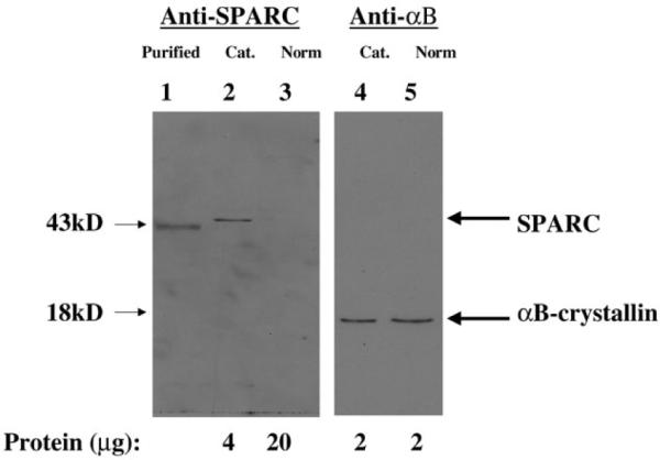
Figure 1.

Western immunoblotting of osteonectin/SPARC in lens extracts. Extracts were prepared and polypeptides were separated and blotted as described in Materials and Methods. Shown is the autoradiogram of the corresponding blot. Lane 1 contains purified osteonectin/SPARC, lane 2 contains cataractous lens extract (cat), and lane 3 contains normal lens extract (norm) probed with anti-SPARC antibody. Lane 4 contains cataractous lens extract (cat) and lane 5 contains normal lens extract (norm) probed with anti-αB-crystallin antibody. Also shown are the osteonectin/SPARC and the αB-crystallin bands with their corresponding molecular weights. The protein added to each well is shown below the blot.
