Figure 3.

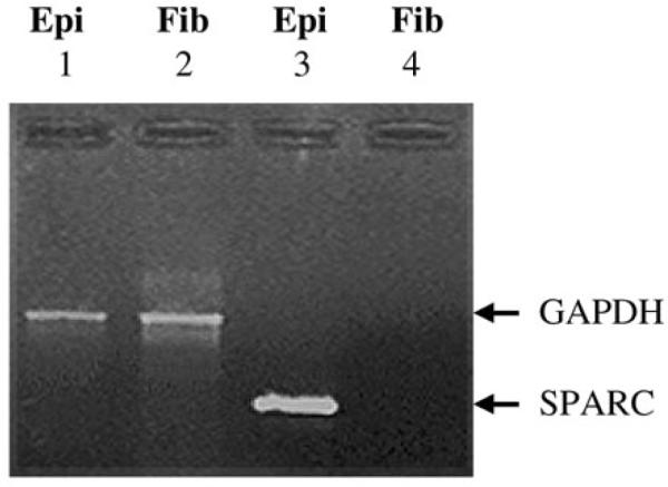
RT-PCR analysis of osteonectin/SPARC in specific cataracts. Ethidium bromide stained gel showing the levels of osteonectin/SPARC and GAPDH in microdissected normal lens epithelia (0.5 μg) and fiber cells (0.5 μg) after 35 PCR cycles. GAPDH transcript (600 bp) levels for lens epithelia (Epi) and fiber cells (Fib) are shown in lanes 1 and 2, respectively. Osteonectin/SPARC transcript (419 bp) levels for lens epithelia (Epi) and fiber cells (Fib) are shown in lanes 3 and 4, respectively.
