
Figure 3.
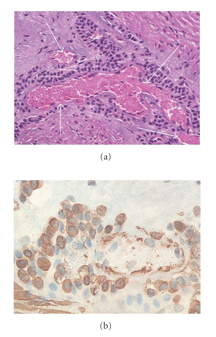
(a) High-magnification hematoxylin (×100) and eosin stained section that demonstrates a fragment of smooth muscle containing infiltrating uniform cells arranged around blood vessels (arrows) and (b) positive immunohistochemical stain for actin which are supportive of a diagnosis of gastric glomus tumor.