
Fig. 2.
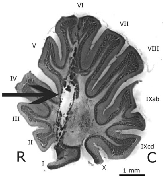
Sagittal section of an adult female zebra finch illustrating a typical lesion (arrow). R, rostral; C, caudal. The lesion tract penetrates folia I–VI and extends into the medial deep cerebellar nucleus. All lesions were bilateral; only a single lesion is visible in this sagittal section. Folia are labeled with roman numerals according to the scheme established by Larsell (1967).