
Fig. 5.
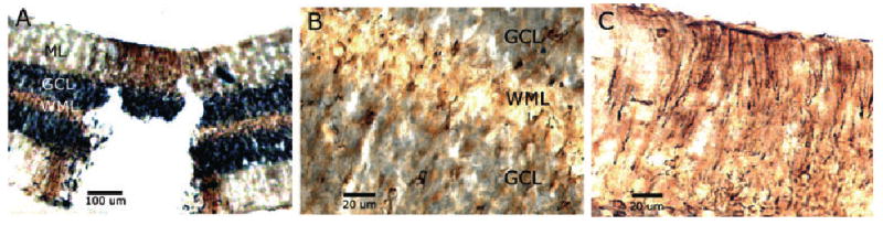
Coronal sections of the cerebellum of an adult female zebra finch showing aromatase immunoreactivity (brown staining) surrounding a cerebellar lesion. (A) Aromatase immunostaining extends laterally from the lesion in the white matter layer (WML) and granule cell layer (GCL) but in the molecular layer (ML) staining occurs only adjacent to the lesion site. (B) Both the GCL and WML show aromatase-positive cells. (C) Aromatase-positive cells in the ML appear to be Bergman glia based on morphology. The GCL stains black non-specifically with this 3′3-diaminobenzidine tetrahydrochloride treatment.