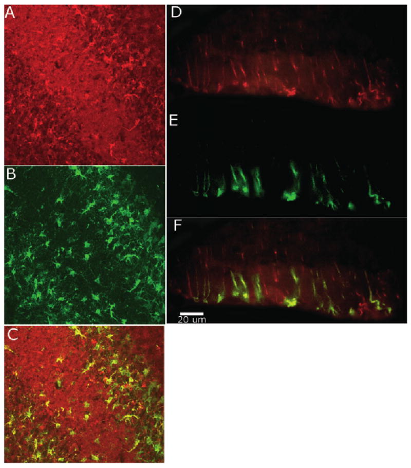
Fig. 6.

Coronal section of injured cerebellum stained for vimentin (Texas red) and aromatase (fluorescein) at 20× (A–C) and 40× (D–F). (A) Vimentin-stained reactive astrocytes in the molecular and white matter layer. (B) Aromatase-stained cells in the molecular and white matter layer. (C) Confocal image identifies considerable overlap between aromatase and vimentin indicating reactive astrocyte aromatase expression. (D) Vimentin-stained Bergmann glia cells in the molecular layer. (E) Aromatase-positive cells in the molecular layer. (F) Overlay of D and E shows Bergmann glia co-expressing vimentin and aromatase.
