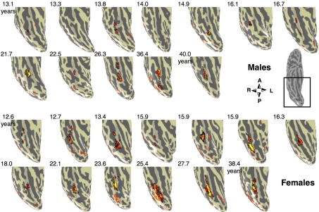
Figure 3.
Face selective activations in the right fusiform gyrus for each of the 25 subjects. Face selective activations for the contrast man and boy > cars and abstract objects, P < 10−3, uncorrected, are projected on the inflated cortical surface of each individual from the right hemisphere for all subjects in the study. Brain images show the posterior aspect of the ventral surface of the right hemisphere as indicated by the inset in a sample brain on the right. Numbers indicate subjects’ age in years. The boundaries of the FFA are shown in black. Compass orients to anterior (A), posterior (P), right (R) or left (L). Top two rows show males’ and bottom rows show females’ brains.

