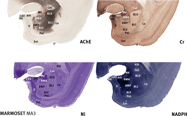
Figure 1.
Marmoset amygdala. Four images of sections taken from the rostrocaudal center of the marmoset amygdala (between 8.0 and 8.5 mm rostral to the interaural plane), each stained with a different marker (Tokuno et al., 2009; see also http://marmoset-brain.org:2008). The dorsal and intermediate part of the basolateral nucleus (BLD, BLI) are clearly defined by intense AChE staining. The basomedial nucleus (BMA and BMP) and the amydalohippocampal area (AHi) are intensely stained with NADPH. The medial nucleus (Me) and the dorsal part of the basomedial nucleus (BMA) are notably stained with Cr. Scale – the field of view in each case is 10-mm wide.

