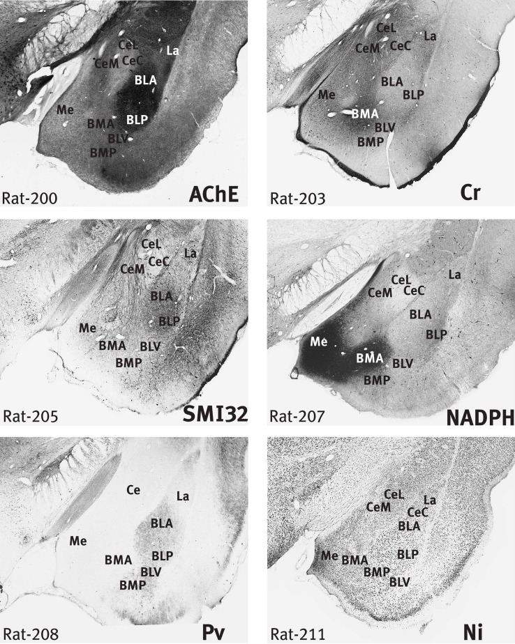
Figure 2.
Rat amygdala. Six images of the rat brain taken from the stained sections used in the preparation of the atlas of Paxinos et al. (2009b), but note that the AChE section is not displayed in the atlas. The sections are numbered from rostral to caudal, and the section thickness is 0.04 mm. The section number is indicated at the lower left. The stain in each case is indicated at the lower right of the section. Scale – the field of view in each case is 4 mm wide. The BLA and BLP are strongly stained in AChE, and the CeC is very lightly stained. Cr staining highlights the BMA and the outer margin of the cortical amygdaloid areas. In NADPH, the Me and BMA are strongly stained, but the CeC is unstained. SMI staining is most marked in the BLA, BLP, and CeM. Pv staining is marked in all parts of BL.

