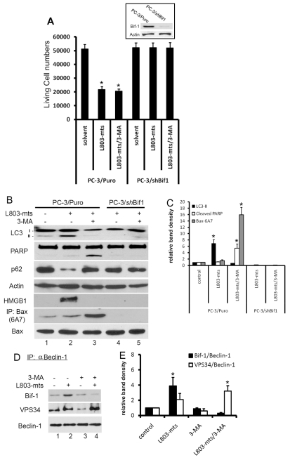
Fig. 6.
Bif-1 is required for GSK-3β-suppression-induced autophagic response and cell death. (A) PC-3/Puro and PC-3/shBif-1 cells were treated with L803-mts (100 μM) with or without 3-MA (10 mM) in serum-free medium for 2 days. Live cells were counted after Trypan Blue staining, as described previously. Inset: exponentially grown PC-3/Puro and PC-3/shBif-1 cells were harvested for western blotting with Bif-1 antibodies. The membrane was re-probed with anti-actin antibody as a loading control. (B,C) PC-3/Puro and PC-3/shBif-1 cells were treated with the solvent or L803-mts (100 μM) plus 3-MA (10 mM) for 16 hours. Cell extracts were subjected to immunoblotting with the antibodies as indicated. To detect Bax conformational change, cellular extracts were prepared in Chaps buffer and then subject to immunoprecipitation with Bax 6A7 antibodies, followed by immunoblotting with regular Bax antibody. Relative band densities were normalized against the anti-actin blot and summarized in C. (D,E) PC-3 cells were treated with L803-mts (100 μM) plus or minus 3-MA (10 mM) in serum-free medium for 18 hours and cellular proteins were subjected to anti-beclin-1 immunoprecipitation and the eluted immunocomplexes were analyzed by western blotting with Bif-1 and VPS34 antibodies, as indicated. Data are from two independent experiments. Relative band densities were shown in E. Asterisks indicate significant difference compared with the control (ANOVA, P<0.05).

